
Following the case study of Phineas Gage scientists, doctors and psychologists started to really investigate the idea that parts of the brain are responsible for certain behaviours and tasks and that any damage to these areas causes abnormalities, like those found in Phineas behaviour after his accident.
If you need a reminder of this case study watch this video
Article about how localisation of the brain has developed over time
The rest of this page covers the following;
- What different parts of the brain are assumed to be related to what behaviours (according to this explanation)
- How to question this explanation of behaviour
- How different hemispheres of the brain are responsible for certain functions
- Research into the division of the two hemisphere’s and how that affects a person’s behaviour
- Evaluating this kind of research
Localisation of the Brain
The theory that specific areas of the brain are associated with particular physical and psychological functions
It is widely recognised that components of the brain can be divided up into the following 3 parts:
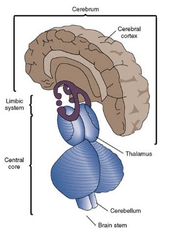
- Central Core
- Limbic System
- Cerebrum
Below describes each of these in a little more detail
Central Core:
The central core describes the five regions of the brain that are present in all vertebrate creatures and are responsible for the most basic and essential biological and sensory life processes.
It is comprised of the thalamus (sensory information processing), medulla (breathing, sleep, heart rate), pons (dreaming, sleep), cerebellum (movement, equilibrium), and reticular formation (stimuli attention, alertness).
You can remember this by using the sentence “The Most Primal Cause and Reason” to recall the first letters of the five regions of the central core.
Limbic System:
The centre for emotional responsiveness, motivation, memory formation and integration (the interaction between different parts of the brain), olfaction(The detection of hazards, pheromones, and food. It integrates with other senses to form the sense of flavour), and the mechanisms to keep ourselves safe.
Cerebrum:
The Cerebrum directs the brains higher cognitive and emotional functions.
It has an outermost layer known as the cerebral cortex; appears grey because of the location of cell bodies (hence “grey-matter”). Each of our sensory systems sends messages to and from this cerebral cortex.
The cerebrum is made up of the left and right hemispheres which are connected by a bundle of fibres called the corpus callosum. This enables messages to enter the right hemisphere to be conveyed to the left hemisphere and vice versa
Difference between grey and white matter for those of you who might be wondering
(you do not need to know this)
The CNS has two kinds of tissue: grey matter and white matter, Grey matter, which has a pinkish-grey colour in the living brain, contains the cell bodies, dendrites and axon terminals of neurons, so it is where all synapses are. White matter is made of axons connecting different parts of grey matter to each other.
For the exam you do not need to know loads of detail about the Central Core and The Limbic system- just a sentence to summarise what it’s role is and be able to name at least 1 part of it. However, The Cerebrum you need to know in much more detail and will be focus of the next part of the page. With all of these parts, The Localisation Explanation suggests that damage to any of these areas would result in the loss of that function.
Parts of the Cerebrum
This part of the brain is the most commonly pictured when someone mentions the brain and is divided up into 4 different lobes. You need to be able to identify where these are in the brain and also what they are responsible. Unfortunately there are so many roles they play that for an A Level qualification it would be unreasonable for the exam board to expect you to be able to identify every last function of every small section of the brain. Therefore we are going to focus on the basics below.
Parietal Lobe: This is named for its proximity to the parietal bone, the wall of the skull. It’s major functions are the primary analysis of somatic sensation (touch, the position of the limbs, temperature) and the analysis of space using all sensory tools. This is called the Somatosensory area. Receptors in different parts of our skin will send information to specific parts of this section. for example, information from our hands and face take up over half of this area because of the amount of receptors we have in them.
Occipital Lobe: (also known as the visual area) Receives and processes visual information. Information from the right-hand side visual field is processed in the left hemisphere, and information from the left-hand side visual field is processed in the right hemisphere. The visual area contains different parts that process different types of information including colour, shape or movement.
Frontal Lobe: This is located at the front of your head, behind your forehead. This tends to be there area of consciousness, decisions making and your personality. towards the back of the frontal lobe is the motor cortex, which is responsible for movement. Damage to the motor cortext would result in lost of function in some or all movement depending on the severity of the damage.
Temporal Lobe: (also known as the auditory area) which analyses speech based information. This area also includes perceiving sounds, assigning meaning to those sounds, and remembering sounds.
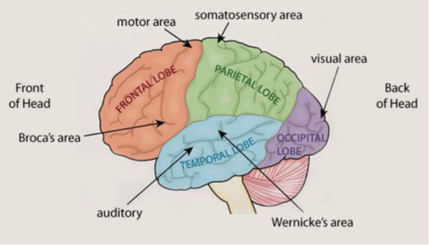
Language Centres: Wernicke and Broca’s Areas
As you will have noticed in the picture above there are two areas you need to know that are called Broca’s and Wernicke’s area. These are responsible for language production and comprehension. It’s very important to remember that these are only in the left side of the brain (Left hemisphere).
Broca’s Area= Speech production. As for the location- think about what you need to be able to speak, movement of your lips and tongue and jaw, but you also need to be able to hear what you are saying as well. Which is why this is found on the boundaries between the frontal lobe (motor cortex) and the temporal lobe (auditory area). This area got it’s name from the French surgeon, Paul Broca in 1861, who found that his patient- nicknames patient Tan- could only say the syllable “Tan”. When he conducted a post-mortem on this person (*AO3 ALERT*) He found damage to this specific part of the brain. It was concluded that this area was responsible for this function and Broca’s Aphasia is a term given to anyone suffering from varying levels of speech impediments. This can range from an inability to speak at all, to only being able to say certain words or syllables.
Video of an man with severe Broca Aphasia
As you can see in the video above, this person can understand what is being said to him, but simply cannot form or get the words out of his mouth. Almost that feeling of having a word on the tip of your tongue but your just can’t say it.
Wernicke= Language comprehension. This is a separate area of language processing which seems to have a specific function. In 1874, Karl Wernicke, who worked at a hospital in Germany, found that patients who had damage in an area close to the auditory cortex had specific language impairments. These included the inability to comprehend language and find the work that they need to use. However, Wernicke did notice that these people has fluent speech, when they could access the words quickly. This led Wernicke to suggest that this area is responsible for understanding language and speaking fluently.
Video of a man with Wernicke’s Aphasia
Evaluation of Localisation
It’s really important to remember with biological explanations of behaviour that just because they are biologically based (some might argue more scientific) that they are not always correct. They have limitations just like the psychodynamic, humanistic and behavioural explanations of behaviour. They might just be limited for other reasons. Within the biological explanations you could talk about brain structure (like this theory) you could discuss neural and hormonal causes or you can use genetics as an explanations. Some explanations might even use all 3. Remember this when asked to discuss an biopsychological explanations of behaviour.
*just because it’s biology doesn’t make it correct!*
Generalisability and application questionable- its really important to remember that post mortems involve studying the brain whilst in an inactive state, which means that you can be certain of that activity when the person was alive and well. Also, there could be other causes of this damage which could be related to the cause of death or the decaying of tissue after death. Additionally, in modern times, with the use of technology, FMRI’s are still not the most ideal method for measuring the brain and establishing a true line of causation between function and localisation. There are so many other factors in an environment that could contribute to activity in the brain that any conclusions about localisation are indirect and could be attributed to other extraneous variables affecting the results. Therefore, Localisation of the brain is explanation that is only loosely support by research, due the to lack of scientifically robust and valid measures to base conclusions on.
Reductionist suggested by Lashley research – Karl Lashley (1950) conducted an experiment on rats where he removed 10-50% of the rats cerebrum when they were learning their way through a maze. He found that it didn’t matter which part of the brain was removed, as it was the amount of the brain that was removed which affects the rats ability work their way around the maze, rather than a specific part. They appeared to need the whole of the cerebrum to complete the task. He suggested that higher cognitive functions, such as learning, are not localised but distributed holistically in the brain.
Reductionist suggested by theory of Plasticity- Another concept that you will learn about later in biopsychology is the idea that the brain can change adapt to ensure specific functions are not lost when trauma or damage happens to particular functions. It works by the brain re-organising itself, by either reconnecting neural pathways, creating brand new neural pathways or moving the function of one part of the brain and redirecting it to another part of the brain. This suggests that localisation of the brain firstly doesn’t accommodate for individual differences, as it might be that due typical wear and tear of life, that each individuals brain might function slightly differently. One persons motor cortex might work or be localised slightly different from the next persons. The fact that localisation of the brain attempts to generalise parts and functioning of the brain to every person in the same way suggests that it is biologically reductionist as it suggest that every person’s brain is made up the same and also that damage to this is irreparable, which has been found to be untrue.
Reductionist (more?!)- Furthermore, psychologists suggest that it is more important to investigate how the brain areas communicate with each other, rather than focusing on specific brain regions. Wernicke claimed that although the different areas of the brain are independent, they must interact with each other in order to function. An example to demonstrate this is a man who lost his ability to read, following damage to the connection between the visual cortex and the Wernicke’s area, which was reported by Dejerine. This suggests that interactions between different areas produce complex behaviours such as language. Therefore, damage to the connection between any two points can result in impairments that resemble damage to the localised brain region associated with that specific function. This reduces the credibility of the localisation theory.
Hemispheric Lateralisation
The dominance of one hemisphere of the brain for particular physical and psychological functions
It is generally assumed, as mentioned above that not only is behaviour and function divided up by the different lobes of the brain but also the different sides. The most widely assumed difference between the left and right is that the left hemisphere is responsible for dealing with language and the right for dealing with spatial awareness. This is why the Broca and Wenicke areas are found in the left hand side of the brain. Identifying which is your left and which is your right is always from your perspective. So your left hand side is your left hemisphere, and your right is your right hemisphere, as demonstrated in the picture below. Her left hemisphere is her left, but would of course be your right- due to the different perspective.
Another difference between the two hemispheres, is that the right side of your brain controls the left side of your body and vice versa. Imagine it like a cross going from one side to the other and back again.

Of course each side of your brain doesn’t work independently of each other, they still need to work together even though the process and activate different information. Think about it, if someone shuts your hand left hand in a car door, that information will be sent to your right hemisphere. What would someone’s reaction be if that happened? Scream or shout from the pain. But your left hemisphere controls language, so how would you react like that if your hemispheres worked completely separate from each other? You couldn’t.
To connect these two side of the brain we have something called the Corpus Callosum. These are a bunch of nerve fibres which run from one side of the brain to the other and back again. Their sole function is to ensure information from one side of the brain is received by the other so that they body and brain can function as one whole unit. Because this transferring of information is happening so consistently and rapidly, like the rest of the brain, it is very difficult in a typical person to be able to pinpoint exactly what information each side if responsible for. You’re probably thinking there much have been some research conducted which supports this theory though otherwise no one would believe it, right?
Split Brain Research
Introducing split brain patients! These are people who, due to extreme epileptic seizures in one side of the brain, have had their corpus callosum cut to stop the electrical activity from one side spreading into the other side of the brain. Your probably now thinking, surely this person now has some form of brain damage and cannot function as a ‘normal’ human being?! Who would be mad enough to 1. suggest it to a person and 2. agree to it?!?!
The research Sperry conducted on his split brain patients is one you need to know really well, as you could be given a 16 or 8 marker on this particular research. Below is details of the procedure and the findings of his study. Whilst you are making notes on this, think about the strengths and weaknesses of this research.
Sperry 1967/8
Aim: The aim of their research was to examine the extent to which the two hemispheres are specialised for certain functions.
Method: An image/word is projected to the patient’s left visual field (which is processed by the right hemisphere) or the right visual field (which is processed by the left hemisphere). When information is presented to one hemisphere in a split-brain patient, the information is not transferred to the other hemisphere (as the corpus callosum is cut).
Sperry and Gazzaniga conducted many different experiments, including describe what you see tasks, tactile tests, and drawing tasks.
In the describe what you see task, a picture was presented to either the left or right visual field and the participant had to simply describe what they saw.
In the tactile test, an object was placed in the patient’s left or right hand and they had to either describe what they felt, or select a similar object from a series of alternate objects.
Finally, in the drawing task, participants were presented with a picture in either their left or right visual field, and they had to simply draw what they saw.
Results:

Conclusion: The findings of Sperry and Gazzaniga’s research highlights a number of key differences between the two hemispheres. Firstly, the left hemisphere is dominant in terms of speech and language. Secondly, the right hemisphere is dominant in terms of visual-motor tasks.
Evaluating Split Brain Research
Generalisability – His sample was a small group of people (11 in total) on a very specific condition. Which means it is a case study. What problems can you think of that would affect our interpretation of the results? Also below is video documenting a person who never developed his corpus callosum. How does his experiences affects how we use and interpret Sperry’s findings?
Validity – Why does conducting a highly controlled lab experiment mean we have low external (mostly ecological validity)? We cannot be certain that they behaviours that occur within this highly controlled lab would occur in exactly the same way out in the real world. Therefore the results Sperry has found can only be applicable within the research setting, an cannot be assumed to be correct in other social settings or environments.
Affects of Plasticity (reductionist?)- By being aware of differences between someone who has had their corpus callosum cut and someone who never developed it, might be evidence of plasticity of the brain. This is the idea that the brain can mould and adapt itself to change based on any damage to it. You will learn about this in more detail on the page labelled “Plasticity and Functional Recovery” . These differences in behavioural symptoms suggest that there is more to the brain than just damaging a part and that actually individual differences as a result of the extent of the damage, the environmental stimuli and how an individuals brain adjusts to change. Something Sperry’s findings from his 11 participants doesn’t account for.
Evaluation of Hemispheric Lateralisation
There is research to support the division of functions between the left and right hemisphere. For example, split brain research, Brocas patient Tan and Wernicke’s research.
Use of scientific measures and lab experiments to test the Lateralisation of hemisphere’s mean it’s can be scientifically tested. For example, the use of post mortems and FMRi’s along with participants such as split brain patients allows for a clear picture of which side of the brain is being used for specific tasks. These methods make use of the empirical method and objective data to make conclusions about brain functions.
The alternative explanation of plasticity draws into question the extent of Lateralisation in the brain. For example, the case study of Jody who, after having half her brain removed, was able to regain many of the functions lost. For example, the movement in the left side of the body is controlled by the right side of the brain. Once this was removed, through recruitment of homologous areas Jody’s brain was able to regain the function that was initially lost.
The usefulness of understanding Hemispheric lateralisation has lead to the development of effective treatments for epilepsy. For example, removing the corpus callosum or in some extreme cases, removing the right side of the brain. This wouldn’t have even been considered without understanding the role of each side.
A weakness of this explanations is it’s reductionist perspective of brain functioning. It simplifies complex brain processes into a clear distinction between the left and right hemispheres, ignoring the intricate networks and interactions within the brain. For instance, language is often associated with the left hemisphere, while creativity with the right. However, this oversimplification disregards the fact that both hemispheres collaborate extensively for most tasks.
This explanation takes a nature perspective, however evidence shows that nurture can also play a role. Hee Yeon (2018) found different hemispheric lateralization patterns: American participants demonstrated a dominance in the right visual field when processing crowd emotion of both European American and Korean facial crowds, and were quicker at recognizing which people to avoid, based on their facial expressions. Whereas, Korean participants showed weak or nonexistent hemisphere dominance. This work suggests that culture plays a role in modulating how our hemisphere’s divide up emotional processing and responses.
