
As you know, the nervous system has two main components. The peripheral nervous system is a communication system, carrying inputs (eg messages from your senses) and outputs (eg messages to tell your muscles to move). Your central nervous system is the data processing system: it processes all the inputs and works out what your responses are going to be, then sends the output message. The whole system is made of neurons which communicate via synapses. So it’s important that we learn about neurons and synapses.
Key definitions
Neuron: A specialised cell which carries signals in the nervous system
Neurotransmitter: A chemical which transmits a signal from one neuron to the next
Synapse: The gap between two neurons
For the exam you need to know the following
- The basic structure of a neuron
- The different types of neurons, how they are structurally different and what their different roles are in the transporting of messages
- Exactly how information passes from one neuron to the next
All of this is covered throughout the rest of this page.
This is a very science heavy part of the spec and requires you to know a lot of key terms, understand them, be able to recall them and them apply that knowledge to unknown situations. The best way to do this is to draw as many diagrams as you can to get your head around it and practice describing and identifying the different key terms and words as often as you can.
Structure of a neuron
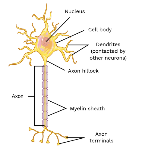
From top to bottom: the dendrites at the top would all have synaptic connections from lots of other neurons. An electrical impulse would be sent along the axon, top to bottom, to the axon terminals. The axon terminals would all have synaptic connections with dendrites of other neurons. Thus neural pathways are formed. The last neuron in the pathway connects to a muscle or gland and the message is actioned.
Below is a description of the role of each part of the neuron;
Cell body: This is the part of the cell that contains the nucleus with its genetic information.
Axon: An axon is a long, tail-like structure which joins the cell body at a specialized junction called the axon hillock. Many axons are insulated with a fatty substance called myelin. Neurons generally have one main axon.
Dendrites: Dendrites branch out from the cell body. Like antennae, dendrites receive and process signals from the axons of other neurons. Neurons can have more than one set of dendrites, known as dendritic trees. How many they have generally depends on their role.
Myelin sheath: Myelin is a fatty substance that acts an electrical insulator. This electrical insulation also speeds up the transmission of the electrical signal.
Nodes of Ranvier: Periodic gaps in the insulating sheath (myelin) on the axon of certain neurons that serves to facilitate the rapid conduction of nerve impulses.
Terminal buttons / axon terminals / presynaptic knobs / presynaptic regions: the small bulbous ends of an axon. When an electrical signal arrives, they release chemicals called neurotransmitters.
Sensory neuron
These neurons provide ‘input’ from the rest of your body to your central nervous system – information from your senses (eyes, ears, pressure, temperature, balance, etc) about your external and internal environments.
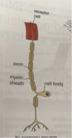
Notice two things about this neuron. (1) It doesn’t have dendrites – instead, it has some kind of sensory receptor, for example pressure receptors in the skin. This is where all the sensory information is collected and sent to the CNS. (2) Its cell body is off to one side, out of the way, so that the electrical impulse can be conducted as fast as possible straight to the CNS.
Motor neuron
This neuron has its cell body in the CNS, and its axon projects outside the CNS. The message will go to a voluntary muscle, an involuntary muscle, or a gland. These are called effector organs. This is how the CNS controls the rest of your body. For example, motor neurons make your muscles contract and relax, so that you can move.
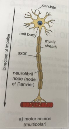
You may have noticed that the terminal buttons of the motor neuron are closely connected to something – this is because its role is to change the activity of a muscle or gland. To do this it needs to communicate directly with that muscle or gland.
Relay neuron / interneuron
These carry messages from one part of the CNS to another. They do not project outside the CNS – ie they are found only in the spinal cord and the brain.
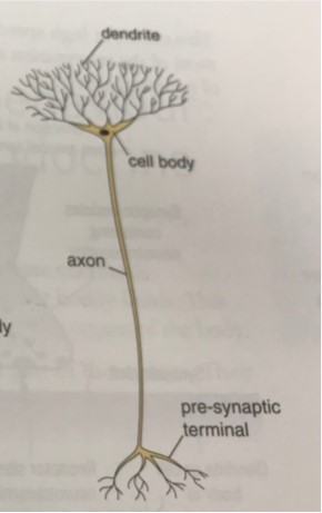
You might have noticed that this is very different from the other two neurons. (1) It has complex branches at each end. This allows for complex interconnections and neural pathways within the CNS. (2) It does not have any myelin or nodes of Ranvier. Remember – myelin’s function is to increase the speed of electrical impulses. Relay neurons don’t need myelin because they are short cells, so their signals will only need to be carried a short distance within the CNS, therefore they don’t need myelination to speed up their signals.
Working together
Click this link to open a YouTube clip talking you through how they all work together
It is important to acknowledge that these are not the only three neuron types that exist in your body – there are subtypes too, so this is a slightly simplified explanation. If this is something that you find interesting, it is definitely worth doing some of your own research and maybe reading some books on it, however for the purpose of the course I think it’s wise to stop there so I can ensure you all have a good grasp of the neurons you need to know!
Here is a link to book titles which you might find satisfy your intellectual curiosity about neurons
Synaptic transmission
So now you know all about neurons, what they are and what they do – now we need to look at how they pass information from one to the other. This is what we call synaptic transmission and requires us to get another magnifying glass out and look even closer at the process going on between each neuron.
Above you will have noticed that each terminal button of one neuron is close to but doesn’t quite touch the dendrites of the next. And if you think about this makes sense – in an electrical circuit, if all the wires physically touched, without any switches, you would have no control over where the signal goes. It’s the same in a neural pathway – if all the neurones touched then all the electrical signals would spread everywhere through the branching cells. Synapses prevent this. Because of synapses, the transmission of neural signals can be controlled. In addition, new synaptic connections can form – this is how learning can take place.
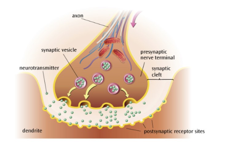
The basics: An electrical signal arrives at the end of the presynaptic neuron. This causes the presynaptic neurone to release its neurotransmitters. Neurotransmitters diffuse across the cleft. When they get to the postsynaptic neurone they will bind there. The postsynaptic neuron will thus have received the signal. It may or may not generate a new electrical impulse.
Below is a picture of this connection in detail all of which you would need to be able to label and describe.
Click on this link to open another YouTube clip describing this process in detail
Exam question: Outline the structures and processes involved in synaptic transmission. (6)
Information is carried by neurons as electrical impulses (sometimes called action potentials). These electrical impulses travel the length of the neuron, all the way along its axon to its branching ends, which are called axon terminals. In a functional nervous system, information needs to pass between neurones, but each neurone does not physically touch the next – there is a gap between neurons called a synapse. Synaptic transmission is how neurons communicate. The presynaptic neuron contains vesicles (sacs) full of chemical neurotransmitters, next to the presynaptic membrane. These are just stored and do not do anything until an electrical impulse reaches the axon terminal. Then, the neurotransmitters are released into the synaptic cleft. The neurotransmitters diffuse across the synapse and bind to receptors on the postsynaptic membrane. This changes the postsynaptic membrane in some way – either by exciting it (eg dopamine), to encourage it to generate a new action potential, or by inhibiting it (eg serotonin), to make it less likely to generate a new impulse.
Excitation and inhibition
To make a working nervous system, only two forces are necessary: excitation and inhibition. Excitatory signaling from one cell to the next makes the latter cell more likely to fire. Inhibitory signalling makes the latter cell less likely to fire. Why do we need two forces? Well, sometimes the CNS needs to create a response, and sometimes it needs to suppress a response.
How are excitation and inhibition involved in synaptic transmission:
- A postsynaptic neuron has lots of dendrites: it has synaptic connections from lots of different presynaptic neurons
- Every synapse releases one type of neurotransmitter only – either excitatory or inhibitory.
- If the synapse’s neurotransmitter is excitatory, then the post synaptic neuron is more likely to fire an impulse.
- If the neurotransmitter is inhibitory then the post synaptic neuron is less likely to fire an impulse.
- The excitatory and inhibitory influences are summed. Consider this situation: a neuron has nine other neurons converging on it. They won’t all be active at once. But, say seven of those synapses are active: if five of the seven are excitatory and two of them are inhibitory, the postsynaptic neuron will generate a new electrical impulse, and send an impulse further along the pathway. If, on the other hand there are six synapses active and four of those are inhibitory – the postsynaptic neuron will not fire.
- The postsynaptic neuron will only generate an impulse if excitation > inhibition.
- Vocab/phrasing: Whether the postsynaptic neuron generates a new impulse is determined by the summation of inhibitory and excitatory signals it is receiving – if overall it is excitatory then an electrical impulse will be generated.
The balance between neural excitation and neural inhibition is crucial to healthy cognition and behaviour. A brain dominated by excitation would only be capable of exciting itself in repeated bursts of activity, similar to an epileptic seizure. A brain dominated by inhibition would only be capable of quiet whispers of activity, with little synchronization necessary for meaningful communication between brain areas.
A simplistic example to illustrate: coughing. You only cough if signals are sent along motor neurons from the CNS to the chest muscles. You may have a few excitatory messages from your airways – there is a tickle, you need to cough. You may also have inhibitory messages from higher areas of the brain – don’t cough, people will think you have covid, or don’t cough because you’re in an exam, or don’t cough because last time you made a weird noise, or don’t cough get a drink instead. If inhibition messages (don’t cough) outweigh excitation messages (do cough), you will suppress the response and motor neurones will not send a ‘cough’ output to the chest muscles. However if your airways get more and more irritated there will be lots more excitatory messages. Once excitation outweighs inhibition, you will cough.
 CGS Psychology Blog: Mrs Harris